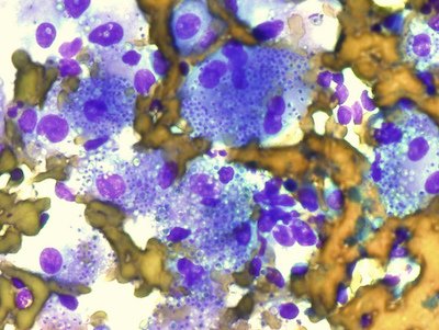
This is histoplasmosis causing respiratory and gastrointestinal signs. Histoplasma capsulatum infection is seen in the river valleys of the central United States, associated with bird and bat droppings in soil.
The big DDx is blastomycosis, also seen in the same area, but with a respiratory presentation. Histoplasmosis is more a chronic diarrheal and respiratory disease in dogs and a respiratory disease in cats. Histoplasma organisms are much smaller than Blastomyces (1-4 μm vs. 8-25 μm) and are difficult to detect with routine H&E stain, unlike Blastomyces organisms.
Use special stains to see yeast forms in macrophages and giant cells (round to ovoid structures, thin cell wall, thin, clear zone between cell wall and cytoplasm). If cytology is not useful consider antigen testing of urine, serum, or CSF. Cross-reactivity with blastomycosis may occur.
Think of coccidioidomycosis in the arid and semiarid southwestern U.S., Mexico, and Central America. Spores are carried on dust and inhaled. Epidemics may occur after dust storms or excavation. Organisms vary in size (20 – 200 μm) and appear as spherules with a double-contoured wall. Mature spherules (sporangia) contain tiny endospores (sporangiospores).
Aspergillosis most commonly causes nasal signs in dogs, such as epistaxis, nasal congestion, and depigmentation of the nasal planum. Disseminated aspergillosis is most commonly seen in middle-aged German shepherds and may cause neurologic signs, lymphadenopathy, and discospondylitis. The organisms appear as narrow, hyaline, septate, branching hyphae on cytology.
Think of cryptococcus in cats with a granulomatous rhinitis and sinusitis. Disseminated disease in dogs and cats may cause central nervous system signs, respiratory signs, or fungal osteomyelitis.
Image courtesy of Yale Rosen.

