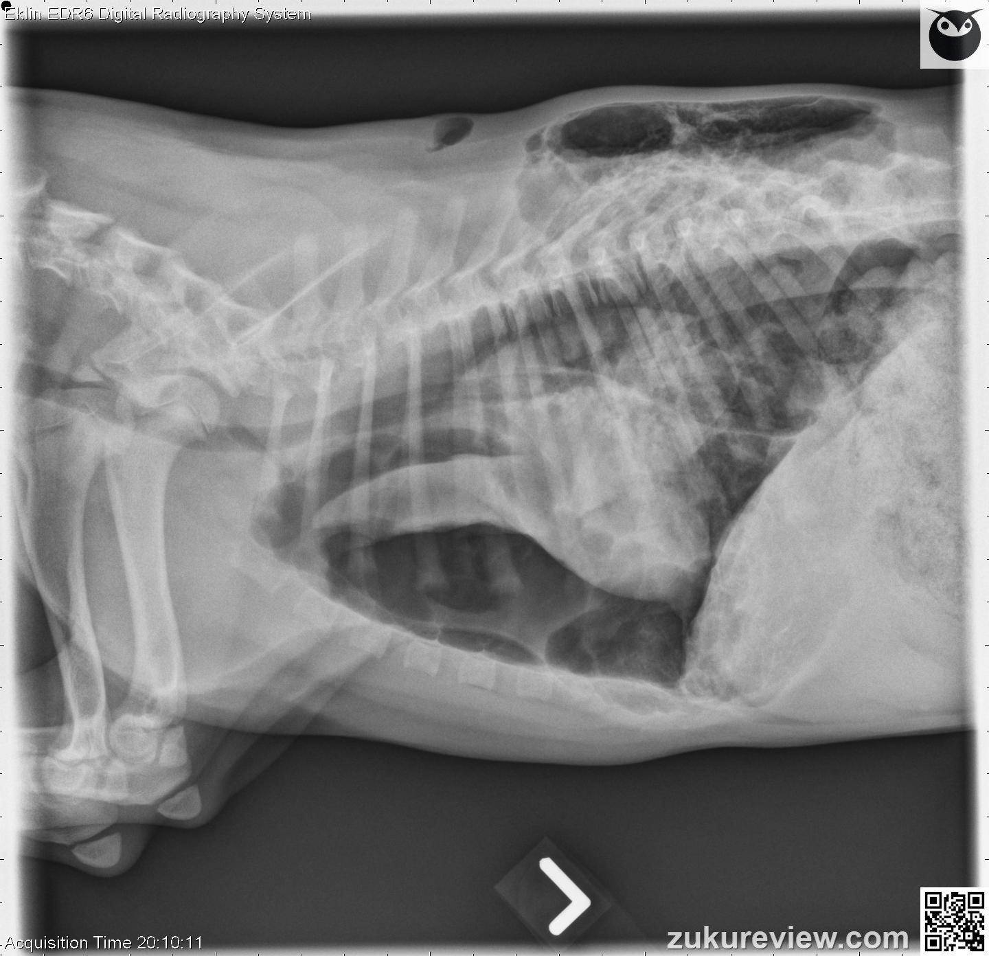
This puppy has a pneumothorax and needs immediate thoracocentesis and chest tube placement. The left hemithorax appears gas distended with marked rightward mediastinal shift.
A large, loculated area of subcutaneous emphysema is seen over the caudal dorsolateral thorax; however, a communication with the thoracic cavity or the external body wall cannot be identified.
A triangular wedge of soft tissue opacity is seen in the mid left thorax and is consistent with collapsed left lung.
A smaller, poorly demarcated area of increased soft tissue opacity is also seen superimposed over the 10th left rib adjacent to the body wall at the level of the most severe subcutaneous emphysema. There is mild pleural effusion in the right hemithorax.
The right pulmonary parenchyma is difficult to evaluate, but appears to be within normal limits. The musculoskeletal structures appear within normal limits.
This is severe left sided tension pneumothorax with collapse of the left lung lobes.
Click here to see normal canine thoracic radiographs
Radiographic interpretation and images courtesy, Dr A. Zwingenberger and Veterinary Radiology. Normal radiograph links courtesy, Imaging Anatomy Univ. of Illinois Vet Med.


