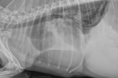
This patient has pleural effusion so perform a thoracocentesis to relieve dyspnea. Fluid analysis and cytology help reach a diagnosis. Ultrasound can guide thoracocentesis but is not strictly necessary.
Causes of pleural effusion in dogs include neoplasia, hemorrhage, idiopathic chylothorax, pyothorax, lung lobe torsion, trauma, and right-sided heart failure.
Radiographic changes associated with pleural effusion include increased soft tissue density in the ventral chest with rounded lung lobe margins and obscuration of the normal cardiac silhouette. Widened interlobar fissure lines are consistent with pleural fluid accumulation.
In this radiograph the lung lobes are displaced dorsally and there is a diffuse interstitial to alveolar pattern, likely as a result of atelectasis secondary to fluid accumulation in the space lungs normally occupy.
Oxygen administration has limited utility as long as the lungs are collapsed from pleural effusion, so prioritize a thoracocentesis over placing nasal oxygen lines. Do not administer antibiotics, beta-blockers, and furosemide until there is more diagnostic data.
Image courtesy of Dr. Teri Defrancesco.

