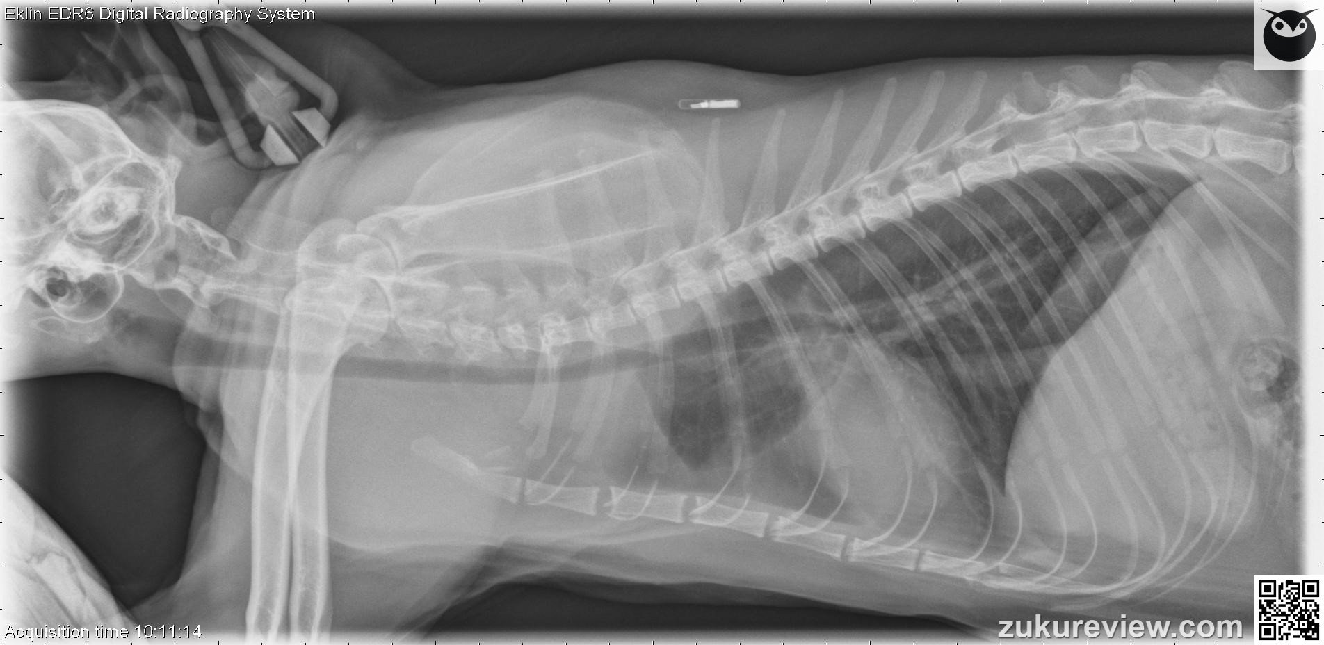
Where is the location of the main lesion in this cat? (Click the images to enlarge)
Correct. You chose {selectedText}.
Incorrect. You chose {selectedText}, but the correct answer is {correctText}.
This cat has a cranial mediastinal mass. There is a small volume pleural effusion causing widened fissure lines and retraction of the lung lobes.
There is focal widening of the cranial mediastinum on the ventrodorsal projection, which is apparent as a soft tissue opacity flattening the margin of the cranial lung lobe on the lateral projection. The trachea is mildly elevated and narrowed in this area.
The most common causes of a cranial mediastinal mass in cats are thymoma and thymic lymphoma. This cat was diagnosed with lymphoma. Rule out FeLV in cats with this condition.
Radiographic interpretation and images courtesy, Dr A. Zwingenberger and Veterinary Radiology. Normal radiograph links courtesy, Imaging Anatomy Univ. of Illinois Vet Med.


