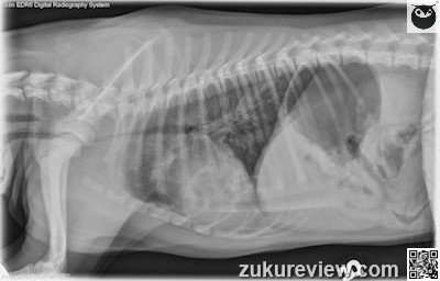
Bronchoscopy is the next best step. The radiographs show an alveolar pulmonary pattern; tracheal narrowing and intraluminal opacity that may indicate edema, mucus, or foreign material; and a dilated pharynx and aerophagia that support an upper airway obstruction.
In more detail: there is an alveolar pulmonary pattern in the left cranial and right middle lung lobes, consistent with aspiration pneumonia, and patchy increased opacity in the remaining lobes (seen best on the left), most consistent w/ non-cardiogenic pulmonary edema.
The trachea is narrowed at the thoracic inlet, with an apparent linear intraluminal opacity. There is dilation of the pharynx with air, and gas within the esophagus and gastrointestinal tract – a common finding in patients w/ respiratory distress from upper airway obstruction.
The dog received a diagnosis of bronchopneumonia, and a seven-inch tracheal foreign body.
Click here to see normal canine thoracic radiographs.
Radiographic interpretation and images courtesy, Dr A. Zwingenberger and Veterinary Radiology. Normal radiograph links courtesy, Imaging Anatomy Univ. of Illinois Vet Med.

