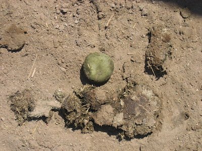
Recommend abdominal radiographs because there is an enterolith in the manure. There is often more than one enterolith, and abdominal radiographs are the least invasive way to evaluate for more (80% sensitivity).
Exploratory laparotomy is the definitive method but is invasive, costly, and not warranted without radiographs and/or clinical signs.
Click here to see an abdominal radiograph from a horse with an enterolith.
Enteroliths are composed of magnesium, ammonium, and phosphate and typically form around a nidus of sand or foreign material. They are most common in Arabians that eat alfalfa hay in certain regions of the USA (CA, southwest, Texas, FL).
While small stones can be passed, enteroliths more often become too large to pass per rectum. They cause recurrent colic or acute colic when they become lodged in narrow bowel (e.g., transverse colon, small colon).
Definitive Tx is ventral midline celiotomy to remove the stones via enterotomy. Prognosis is excellent following surgical removal, although there are the same risks as for all exploratory laparotomies (risk of anesthesia, incisional infection, etc.).
Images courtesy of Nora Grenager, VMD, DACVIM.

