A six-year-old miniature Dachshund is presented with progressively worsening carpal and tarsal laxity.
Which one of the following choices is the most likely diagnosis?
Correct. You chose {selectedText}.
Incorrect. You chose {selectedText}, but the correct answer is {correctText}.
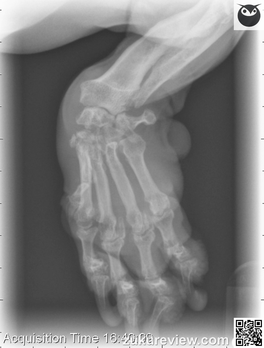
This dog has rheumatoid arthritis (erosive polyarthritis). There is mild to moderate destruction of the cortex of the distal radius.
There is decreased mineral opacity in the distal carpal bones and large lucent areas are visible within them. The cortices of the distal carpal bones are not well visualized.
The cortical margins of the carpal bones are irregular. The proximal and distal intercarpal joints are collapsed.
There is increased intracapsular soft tissue opacity in the carpal joint. There is a moderate soft tissue swelling at the level of the carpal joint.
Radiographic interpretation and images courtesy, Dr A. Zwingenberger and Veterinary Radiology. Normal radiograph links courtesy, Imaging AnatomyUniv. of Illinois Vet Med.


